Human Anatomy and Physiology : Introduction to the Muscular System
Introduction to the Muscular System
The muscular system is composed of specialized cells called muscle fibers. Their predominant function is contractibility. Muscles, attached to bones or internal organs and blood vessels, are responsible for movement. Nearly all movement in the body is the result of muscle contraction. Exceptions to this are the action of cilia, the flagellum on sperm cells, and amoeboid movement of some white blood cells.
The integrated action of joints, bones, and skeletal muscles produces obvious movements such as walking and running. Skeletal muscles also produce more subtle movements that result in various facial expressions, eye movements, and respiration.
In addition to movement, muscle contraction also fulfills some other important functions in the body, such as posture, joint stability, and heat production. Posture, such as sitting and standing, is maintained as a result of muscle contraction. The skeletal muscles are continually making fine adjustments that hold the body in stationary positions. The tendons of many muscles extend over joints and in this way contribute to joint stability. This is particularly evident in the knee and shoulder joints, where muscle tendons are a major factor in stabilizing the joint. Heat production, to maintain body temperature, is an important by-product of muscle metabolism. Nearly 85 percent of the heat produced in the body is the result of muscle contraction.
Structure of Skeletal Muscle
A whole skeletal muscle is considered an organ of the muscular system. Each organ or muscle consists of skeletal muscle tissue, connective tissue, nerve tissue, and blood or vascular tissue.
Skeletal muscles vary considerably in size, shape, and arrangement of fibers. They range from extremely tiny strands such as the stapedium muscle of the middle ear to large masses such as the muscles of the thigh. Some skeletal muscles are broad in shape and some narrow. In some muscles the fibers are parallel to the long axis of the muscle; in some they converge to a narrow attachment; and in some they are oblique.
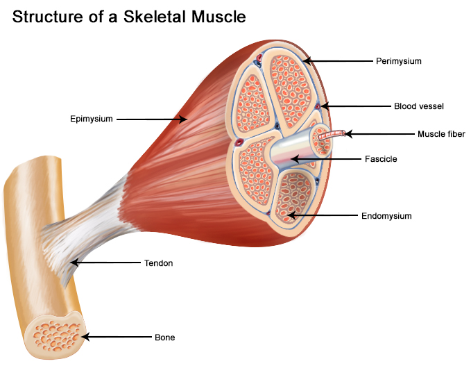
Each skeletal muscle fiber is a single cylindrical muscle cell. An individual skeletal muscle may be made up of hundreds, or even thousands, of muscle fibers bundled together and wrapped in a connective tissue covering. Each muscle is surrounded by a connective tissue sheath called the epimysium. Fascia, connective tissue outside the epimysium, surrounds and separates the muscles. Portions of the epimysium project inward to divide the muscle into compartments. Each compartment contains a bundle of muscle fibers. Each bundle of muscle fiber is called a fasciculus and is surrounded by a layer of connective tissue called the perimysium. Within the fasciculus, each individual muscle cell, called a muscle fiber, is surrounded by connective tissue called the endomysium.
Skeletal muscle cells (fibers), like other body cells, are soft and fragile. The connective tissue covering furnish support and protection for the delicate cells and allow them to withstand the forces of contraction. The coverings also provide pathways for the passage of blood vessels and nerves.
Commonly, the epimysium, perimysium, and endomysium extend beyond the fleshy part of the muscle, the belly or gaster, to form a thick ropelike tendon or a broad, flat sheet-like aponeurosis. The tendon and aponeurosis form indirect attachments from muscles to the periosteum of bones or to the connective tissue of other muscles. Typically a muscle spans a joint and is attached to bones by tendons at both ends. One of the bones remains relatively fixed or stable while the other end moves as a result of muscle contraction.
Skeletal muscles have an abundant supply of blood vessels and nerves. This is directly related to the primary function of skeletal muscle, contraction. Before a skeletal muscle fiber can contract, it has to receive an impulse from a nerve cell. Generally, an artery and at least one vein accompany each nerve that penetrates the epimysium of a skeletal muscle. Branches of the nerve and blood vessels follow the connective tissue components of the muscle of a nerve cell and with one or more minute blood vessels called capillaries.
Muscle Types
In the body, there are three types of muscle: skeletal (striated), smooth, and cardiac.
Skeletal Muscle
Skeletal muscle, attached to bones, is responsible for skeletal movements. The peripheral portion of the central nervous system (CNS) controls the skeletal muscles. Thus, these muscles are under conscious, or voluntary, control. The basic unit is the muscle fiber with many nuclei. These muscle fibers are striated (having transverse streaks) and each acts independently of neighboring muscle fibers.
Smooth Muscle
Smooth muscle, found in the walls of the hollow internal organs such as blood vessels, the gastrointestinal tract, bladder, and uterus, is under control of the autonomic nervous system. Smooth muscle cannot be controlled consciously and thus acts involuntarily. The non-striated (smooth) muscle cell is spindle-shaped and has one central nucleus. Smooth muscle contracts slowly and rhythmically.
Cardiac Muscle
Cardiac muscle, found in the walls of the heart, is also under control of the autonomic nervous system. The cardiac muscle cell has one central nucleus, like smooth muscle, but it also is striated, like skeletal muscle. The cardiac muscle cell is rectangular in shape. The contraction of cardiac muscle is involuntary, strong, and rhythmical.
Smooth and cardiac muscle will be discussed in detail with respect to their appropriate systems. This unit mainly covers the skeletal muscular system.
Muscle Groups
There are more than 600 muscles in the body, which together account for about 40 percent of a person's weight.
Most skeletal muscles have names that describe some feature of the muscle. Often several criteria are combined into one name. Associating the muscle's characteristics with its name will help you learn and remember them. The following are some terms relating to muscle features that are used in naming muscles.
- Size: vastus (huge); maximus (large); longus (long); minimus (small); brevis (short).
- Shape: deltoid (triangular); rhomboid (like a rhombus with equal and parallel sides); latissimus (wide); teres (round); trapezius (like a trapezoid, a four-sided figure with two sides parallel).
- Direction of fibers: rectus (straight); transverse (across); oblique (diagonally); orbicularis (circular).
- Location: pectoralis (chest); gluteus (buttock or rump); brachii (arm); supra- (above); infra- (below); sub- (under or beneath); lateralis (lateral).
- Number of origins: biceps (two heads); triceps (three heads); quadriceps (four heads).
- Origin and insertion: sternocleidomastoideus (origin on the sternum and clavicle, insertion on the mastoid process); brachioradialis (origin on the brachium or arm, insertion on the radius).
- Action: abductor (to abduct a structure); adductor (to adduct a structure); flexor (to flex a structure); extensor (to extend a structure); levator (to lift or elevate a structure); masseter (a chewer).
Muscles of the Head and Neck
Humans have well-developed muscles in the face that permit a large variety of facial expressions. Because the muscles are used to show surprise, disgust, anger, fear, and other emotions, they are an important means of nonverbal communication. Muscles of facial expression include frontalis, orbicularis oris, laris oculi, buccinator, and zygomaticus.These muscles of facial expressions are identified in the illustration below.
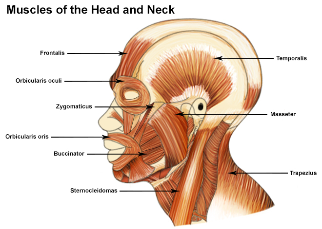
There are four pairs of muscles that are responsible for chewing movements or mastication. All of these muscles connect to the mandible and they are some of the strongest muscles in the body. Two of the muscles, temporalis and masseter, are identified in the illustration above.
There are numerous muscles associated with the throat, the hyoid bone and the vertebral column; only two of the more obvious and superficial neck muscles are identified in the illustration: sternocleidomastoid and trapezius.
Muscles of the Trunk
The muscles of the trunk include those that move the vertebral column, the muscles that form the thoracic and abdominal walls, and those that cover the pelvic outlet.
The erector spinae group of muscles on each side of the vertebral column is a large muscle mass that extends from the sacrum to the skull. These muscles are primarily responsible for extending the vertebral column to maintain erect posture. The deep back muscles occupy the space between the spinous and transverse processes of adjacent vertebrae.
The muscles of the thoracic wall are involved primarily in the process of breathing. The intercostal muscles are located in spaces between the ribs. They contract during forced expiration. External intercostal muscles contract to elevate the ribs during the inspiration phase of breathing. The diaphragm is a dome-shaped muscle that forms a partition between the thorax and the abdomen. It has three openings in it for structures that have to pass from the thorax to the abdomen.
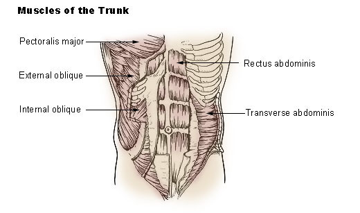
The abdomen, unlike the thorax and pelvis, has no bony reinforcements or protection. The wall consists entirely of four muscle pairs, arranged in layers, and the fascia that envelops them. The abdominal wall muscles are identified in the illustration below.
The pelvic outlet is formed by two muscular sheets and their associated fascia.
Muscles of the Upper Extremity
The muscles of the upper extremity include those that attach the scapula to the thorax and generally move the scapula, those that attach the humerus to the scapula and generally move the arm, and those that are located in the arm or forearm that move the forearm, wrist, and hand. The illustration below shows some of the muscles of the upper extremity.
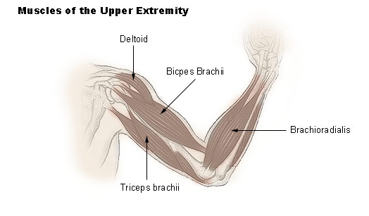
Muscles that move the shoulder and arm include the trapezius and serratus anterior. The pectoralis major, latissimus dorsi, deltoid, and rotator cuff muscles connect to the humerus and move the arm.
The muscles that move the forearm are located along the humerus, which include the triceps brachii, biceps brachii, brachialis, and brachioradialis. The 20 or more muscles that cause most wrist, hand, and finger movements are located along the forearm.
Muscles of the Lower Extremity
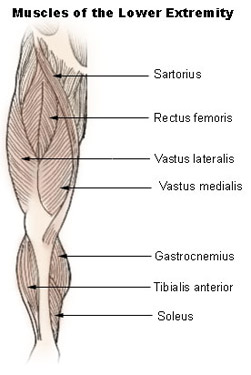
The muscles that move the thigh have their origins on some part of the pelvic girdle and their insertions on the femur. The largest muscle mass belongs to the posterior group, the gluteal muscles, which, as a group, adduct the thigh. The iliopsoas, an anterior muscle, flexes the thigh. The muscles in the medial compartment adduct the thigh. The illustration below shows some of the muscles of the lower extremity.
Muscles that move the leg are located in the thigh region. The quadriceps femoris muscle group straightens the leg at the knee. The hamstrings are antagonists to the quadriceps femoris muscle group, which are used to flex the leg at the knee.
The muscles located in the leg that move the ankle and foot are divided into anterior, posterior, and lateral compartments. The tibialis anterior, which dorsiflexes the foot, is antagonistic to the gastrocnemius and soleus muscles, which plantar flex the foot.
Review: Introduction to the Muscular System
Here is what we have learned from Introduction to the Muscular System:
- One of the most predominant characteristics of skeletal muscle tissue is its contractility and nearly all movement in the body is the result of muscle contraction.
- Four functions of muscle contraction are movement, posture, joint stability, and heat production.
- Three types of muscle are skeletal, smooth, and cardiac.
- Each muscle fiber is surrounded by endomysium. The fibers are collected into bundles covered by perimysium. Many bundles, or fasciculi, are wrapped together by the epimysium to form a whole muscle.
- Muscles are attached to bones by tendons.
- Muscle features such as size, shape, direction of fibers, location, number of origin, origin and insertion, and action are often used in naming muscles.
- Four major muscle groups of the body include:
- Muscles of the head and neck;
- Muscles of the trunk;
- Muscles of the upper extremity; and
- Muscles of the lower extremity.


Comments
Post a Comment