Human Anatomy and Physiology : Introduction to the Nervous System
Introduction to the Nervous System
The nervous system is the major controlling, regulatory, and communicating system in the body. It is the center of all mental activity including thought, learning, and memory. Together with the endocrine system, the nervous system is responsible for regulating and maintaining homeostasis. Through its receptors, the nervous system keeps us in touch with our environment, both external and internal.
Like other systems in the body, the nervous system is composed of organs, principally the brain, spinal cord, nerves, and ganglia. These, in turn, consist of various tissues, including nerve, blood, and connective tissue. Together these carry out the complex activities of the nervous system.
The various activities of the nervous system can be grouped together as three general, overlapping functions:
- Sensory
- Integrative
- Motor
Millions of sensory receptors detect changes, called stimuli, which occur inside and outside the body. They monitor such things as temperature, light, and sound from the external environment. Inside the body, the internal environment, receptors detect variations in pressure, pH, carbon dioxide concentration, and the levels of various electrolytes. All of this gathered information is called sensory input.
Sensory input is converted into electrical signals called nerve impulses that are transmitted to the brain. There the signals are brought together to create sensations, to produce thoughts, or to add to memory; Decisions are made each moment based on the sensory input. This is integration.
Based on the sensory input and integration, the nervous system responds by sending signals to muscles, causing them to contract, or to glands, causing them to produce secretions. Muscles and glands are called effectors because they cause an effect in response to directions from the nervous system. This is the motor output or motor function.
Nerve Tissue
Although the nervous system is very complex, there are only two main types of cells in nerve tissue. The actual nerve cell is the neuron. It is the "conducting" cell that transmits impulses and the structural unit of the nervous system. The other type of cell is neuroglia, or glial, cell. The word "neuroglia" means "nerve glue." These cells are nonconductive and provide a support system for the neurons. They are a special type of "connective tissue" for the nervous system.
Neurons
Neurons, or nerve cells, carry out the functions of the nervous system by conducting nerve impulses. They are highly specialized and amitotic. This means that if a neuron is destroyed, it cannot be replaced because neurons do not go through mitosis. The image below illustrates the structure of a typical neuron.
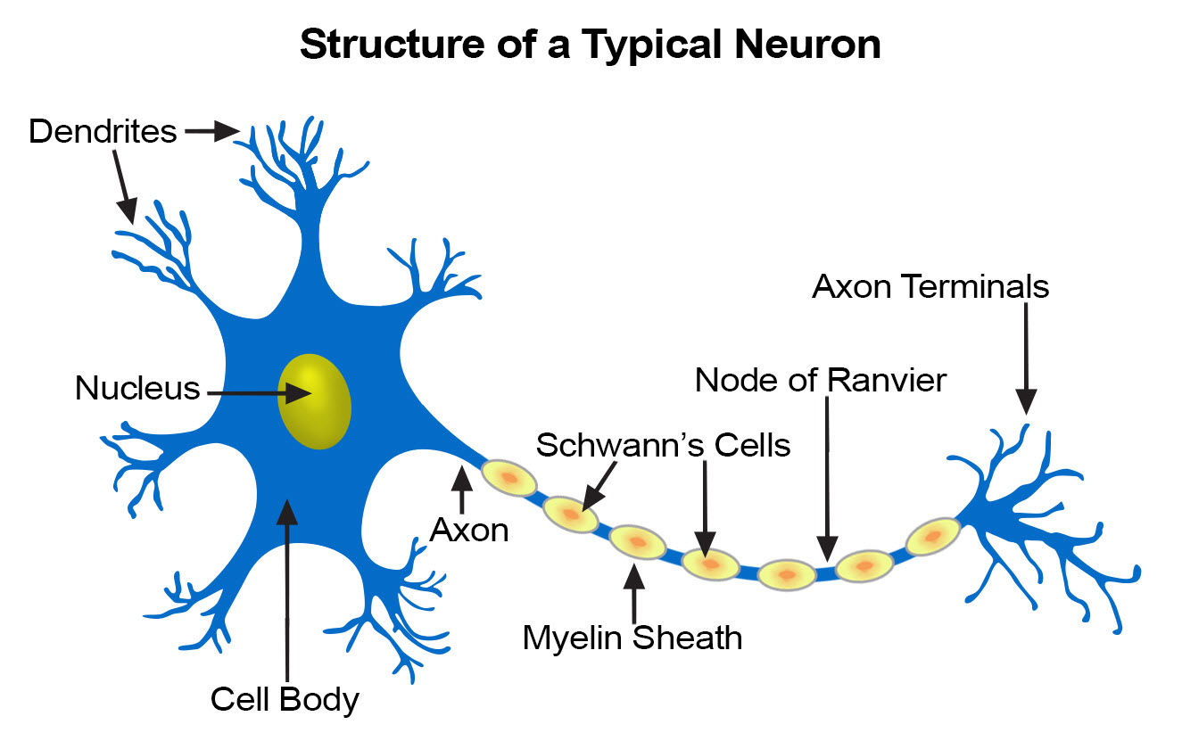
Each neuron has three basic parts: cell body (soma), one or more dendrites, and a single axon.
Cell Body
In many ways, the cell body is similar to other types of cells. It has a nucleus with at least one nucleolus and contains many of the typical cytoplasmic organelles. It lacks centrioles, however. Because centrioles function in cell division, the fact that neurons lack these organelles is consistent with the amitotic nature of the cell.
Dendrites
Dendrites and axons are cytoplasmic extensions, or processes, that project from the cell body. They are sometimes referred to as fibers. Dendrites are usually, but not always, short and branching, which increases their surface area to receive signals from other neurons. The number of dendrites on a neuron varies. They are called afferent processes because they transmit impulses to the neuron cell body. There is only one axon that projects from each cell body. It is usually elongated and because it carries impulses away from the cell body, it is called an efferent process.
Axon
An axon may have infrequent branches called axon collaterals. Axons and axon collaterals terminate in many short branches or telodendria. The distal ends of the telodendria are slightly enlarged to form synaptic bulbs. Many axons are surrounded by a segmented, white, fatty substance called myelin or the myelin sheath. Myelinated fibers make up the white matter in the CNS, while cell bodies and unmyelinated fibers make the gray matter. The unmyelinated regions between the myelin segments are called the nodes of Ranvier.
In the peripheral nervous system, the myelin is produced by Schwann cells. The cytoplasm, nucleus, and outer cell membrane of the Schwann cell form a tight covering around the myelin and around the axon itself at the nodes of Ranvier. This covering is the neurilemma, which plays an important role in the regeneration of nerve fibers. In the CNS, oligodendrocytes produce myelin, but there is no neurilemma, which is why fibers within the CNS do not regenerate.
Functionally, neurons are classified as afferent, efferent, or interneurons (association neurons) according to the direction in which they transmit impulses relative to the central nervous system. Afferent, or sensory, neurons carry impulses from peripheral sense receptors to the CNS. They usually have long dendrites and relatively short axons. Efferent, or motor, neurons transmit impulses from the CNS to effector organs such as muscles and glands. Efferent neurons usually have short dendrites and long axons. Interneurons, or association neurons, are located entirely within the CNS in which they form the connecting link between the afferent and efferent neurons. They have short dendrites and may have either a short or long axon.
Neuroglia
Neuroglia cells do not conduct nerve impulses, but instead, they support, nourish, and protect the neurons. They are far more numerous than neurons and, unlike neurons, are capable of mitosis.
Tumors
Schwannomas are benign tumors of the peripheral nervous system which commonly occur in their sporadic, solitary form in otherwise normal individuals. Rarely, individuals develop multiple schwannomas arising from one or many elements of the peripheral nervous system.
Commonly called a Morton's Neuroma, this problem is a fairly common benign nerve growth and begins when the outer coating of a nerve in your foot thickens. This thickening is caused by irritation of branches of the medial and lateral plantar nerves that results when two bones repeatedly rub together.
Organization of the Nervous System
Although terminology seems to indicate otherwise, there is really only one nervous system in the body. Although each subdivision of the system is also called a "nervous system," all of these smaller systems belong to the single, highly integrated nervous system. Each subdivision has structural and functional characteristics that distinguish it from the others. The nervous system as a whole is divided into two subdivisions: the central nervous system (CNS) and the peripheral nervous system (PNS).
The Central Nervous System
The brain and spinal cord are the organs of the central nervous system. Because they are so vitally important, the brain and spinal cord, located in the dorsal body cavity, are encased in bone for protection. The brain is in the cranial vault, and the spinal cord is in the vertebral canal of the vertebral column. Although considered to be two separate organs, the brain and spinal cord are continuous at the foramen magnum.
The Peripheral Nervous System
The organs of the peripheral nervous system are the nerves and ganglia. Nerves are bundles of nerve fibers, much like muscles are bundles of muscle fibers. Cranial nerves and spinal nerves extend from the CNS to peripheral organs such as muscles and glands. Ganglia are collections, or small knots, of nerve cell bodies outside the CNS.
The peripheral nervous system is further subdivided into an afferent (sensory) division and an efferent (motor) division. The afferent or sensory division transmits impulses from peripheral organs to the CNS. The efferent or motor division transmits impulses from the CNS out to the peripheral organs to cause an effect or action.
Finally, the efferent or motor division is again subdivided into the somatic nervous system and the autonomic nervous system. The somatic nervous system, also called the somatomotor or somatic efferent nervous system, supplies motor impulses to the skeletal muscles. Because these nerves permit conscious control of the skeletal muscles, it is sometimes called the voluntary nervous system. The autonomic nervous system, also called the visceral efferent nervous system, supplies motor impulses to cardiac muscle, to smooth muscle, and to glandular epithelium. It is further subdivided into sympathetic and parasympathetic divisions. Because the autonomic nervous system regulates involuntary or automatic functions, it is called the involuntary nervous system.
The Central Nervous System
The CNS consists of the brain and spinal cord, which are located in the dorsal body cavity. The brain is surrounded by the cranium, and the spinal cord is protected by the vertebrae. The brain is continuous with the spinal cord at the foramen magnum. In addition to bone, the CNS is surrounded by connective tissue membranes, called meninges, and by cerebrospinal fluid.
Meninges
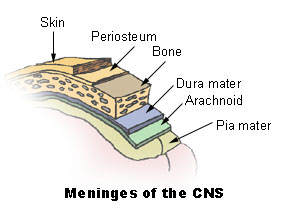
There are three layers of meninges around the brain and spinal cord. The outer layer, the dura mater, is tough white fibrous connective tissue. The middle layer of meninges is arachnoid, which resembles a cobweb in appearance, is a thin layer with numerous threadlike strands that attach it to the innermost layer. The space under the arachnoid, the subarachnoid space, is filled with cerebrospinal fluid and contains blood vessels. The pia mater is the innermost layer of meninges. This thin, delicate membrane is tightly bound to the surface of the brain and spinal cord and cannot be dissected away without damaging the surface.
Meningiomas are tumors of the nerve tissue covering the brain and spinal cord. Although meningiomas are usually not likely to spread, physicians often treat them as though they were malignant to treat symptoms that may develop when a tumor applies pressure to the brain.
Brain
The brain is divided into the cerebrum, diencephalons, brain stem, and cerebellum.
Cerebrum
The largest and most obvious portion of the brain is the cerebrum, which is divided by a deep longitudinal fissure into two cerebral hemispheres. The two hemispheres are two separate entities but are connected by an arching band of white fibers, called the corpus callosum that provides a communication pathway between the two halves.
Each cerebral hemisphere is divided into five lobes, four of which have the same name as the bone over them: the fontal lobe, the parietal lobe, the occipital lobe, and the temporal lobe. A fifth lobe, the insula or Island of Reil, lies deep within the lateral sulcus.
Diencephalon
The diencephalons is centrally located and is nearly surrounded by the cerebral hemispheres. It includes the thalamus, hypothalamus, and epithalamus. The thalamus, about 80 percent of the diencephalons, consists of two oval masses of gray matter that serve as relay stations for sensory impulses, except for the sense of smell, going to the cerebral cortex. The hypothalamus is a small region below the thalamus, which plays a key role in maintaining homeostasis because it regulates many visceral activities. The epithalamus is the most dorsal portion of the diencephalons. This small gland is involved with the onset of puberty and rhythmic cycles in the body. It is like a biological clock.
Brain Stem
The brain stem is the region between the diencephalons and the spinal cord. It consists of three parts: midbrain, pons, and medulla oblongata. The midbrain is the most superior portion of the brain stem. The pons is the bulging middle portion of the brain stem. This region primarily consists of nerve fibers that form conduction tracts between the higher brain centers and spinal cord. The medulla oblongata, or simply medulla, extends inferiorly from the pons. It is continuous with the spinal cord at the foramen magnum. All the ascending (sensory) and descending (motor) nerve fibers connecting the brain and spinal cord pass through the medulla.
Cerebellum
The cerebellum, the second largest portion of the brain, is located below the occipital lobes of the cerebrum. Three paired bundles of myelinated nerve fibers, called cerebellar peduncles, form communication pathways between the cerebellum and other parts of the central nervous system.
Ventricles and Cerebrospinal Fluid
A series of interconnected, fluid-filled cavities are found within the brain. These cavities are the ventricles of the brain, and the fluid is cerebrospinal fluid (CSF).
Spinal Cord
The spinal cord extends from the foramen magnum at the base of the skull to the level of the first lumbar vertebra. The cord is continuous with the medulla oblongata at the foramen magnum. Like the brain, the spinal cord is surrounded by bone, meninges, and cerebrospinal fluid.
The spinal cord is divided into 31 segments with each segment giving rise to a pair of spinal nerves. At the distal end of the cord, many spinal nerves extend beyond the conus medullaris to form a collection that resembles a horse's tail. This is the cauda equina. In cross section, the spinal cord appears oval in shape.
The spinal cord has two main functions:
- Serving as a conduction pathway for impulses going to and from the brain. Sensory impulses travel to the brain on ascending tracts in the cord. Motor impulses travel on descending tracts.
- Serving as a reflex center. The reflex arc is the functional unit of the nervous system. Reflexes are responses to stimuli that do not require conscious thought and consequently, they occur more quickly than reactions that require thought processes. For example, with the withdrawal reflex, the reflex action withdraws the affected part before you are aware of the pain. Many reflexes are mediated in the spinal cord without going to the higher brain centers.
Brain Tumor
Glioma refers to tumors that arise from the support cells of the brain. These cells are called glial cells. These tumors include the astrocytomas, ependymomas and oligodendrogliomas. These tumors are the most common primary brain tumors.
The Peripheral Nervous System
The peripheral nervous system consists of the nerves that branch out from the brain and spinal cord. These nerves form the communication network between the CNS and the body parts. The peripheral nervous system is further subdivided into the somatic nervous system and the autonomic nervous system. The somatic nervous system consists of nerves that go to the skin and muscles and is involved in conscious activities. The autonomic nervous system consists of nerves that connect the CNS to the visceral organs such as the heart, stomach, and intestines. It mediates unconscious activities.
Structure of a Nerve
A nerve contains bundles of nerve fibers, either axons or dendrites, surrounded by connective tissue. Sensory nerves contain only afferent fibers, long dendrites of sensory neurons. Motor nerves have only efferent fibers, long axons of motor neurons. Mixed nerves contain both types of fibers.
A connective tissue sheath called the epineurium surrounds each nerve. Each bundle of nerve fibers is called a fasciculus and is surrounded by a layer of connective tissue called the perineurium. Within the fasciculus, each individual nerve fiber, with its myelin and neurilemma, is surrounded by connective tissue called the endoneurium. A nerve may also have blood vessels enclosed in its connective tissue wrappings.
Cranial Nerves
Twelve pairs of cranial nerves emerge from the inferior surface of the brain. All of these nerves, except the vagus nerve, pass through foramina of the skull to innervate structures in the head, neck, and facial region.
The cranial nerves are designated both by name and by Roman numerals, according to the order in which they appear on the inferior surface of the brain. Most of the nerves have both sensory and motor components. Three of the nerves are associated with the special senses of smell, vision, hearing, and equilibrium and have only sensory fibers. Five other nerves are primarily motor in function but do have some sensory fibers for proprioception. The remaining four nerves consist of significant amounts of both sensory and motor fibers.
Acoustic neuromas are benign fibrous growths that arise from the balance nerve, also called the eighth cranial nerve or vestibulocochlear nerve. These tumors are non-malignant, meaning that they do not spread or metastasize to other parts of the body. The location of these tumors is deep inside the skull, adjacent to vital brain centers in the brain stem. As the tumors enlarge, they involve surrounding structures which have to do with vital functions. In the majority of cases, these tumors grow slowly over a period of years. In other cases, the growth rate is more rapid and patients develop symptoms at a faster pace. Usually, the symptoms are mild and many patients are not diagnosed until some time after their tumor has developed. Many patients also exhibit no tumor growth over a number of years when followed by yearly MRI scans.
Spinal Nerves
Thirty-one pairs of spinal nerves emerge laterally from the spinal cord. Each pair of nerves corresponds to a segment of the cord and they are named accordingly. This means there are 8 cervical nerves, 12 thoracic nerves, 5 lumbar nerves, 5 sacral nerves, and 1 coccygeal nerve.
Each spinal nerve is connected to the spinal cord by a dorsal root and a ventral root. The cell bodies of the sensory neurons are in the dorsal root ganglion, but the motor neuron cell bodies are in the gray matter. The two roots join to form the spinal nerve just before the nerve leaves the vertebral column. Because all spinal nerves have both sensory and motor components, they are all mixed nerves.
Autonomic Nervous System
The autonomic nervous system is a visceral efferent system, which means it sends motor impulses to the visceral organs. It functions automatically and continuously, without conscious effort, to innervate smooth muscle, cardiac muscle, and glands. It is concerned with heart rate, breathing rate, blood pressure, body temperature, and other visceral activities that work together to maintain homeostasis.
The autonomic nervous system has two parts, the sympathetic division and the parasympathetic division. Many visceral organs are supplied with fibers from both divisions. In this case, one stimulates and the other inhibits. This antagonistic functional relationship serves as a balance to help maintain homeostasis.
Review: Introduction to the Nervous System
Here is what we have learned from this Introduction to the Nervous System:
- The nervous system is the major controlling, regulatory, and communicating system in the body. It is the center of all mental activity including thought, learning, and memory.
- The various activities of the nervous system can be grouped together as three general, overlapping functions: sensory, integrative, and motor.
- Neurons are the nerve cells that transmit impulses. Supporting cells are neuroglia.
- The three components of a neuron are a cell body or soma, one or more afferent processes called dendrites, and a single efferent process called an axon.
- The central nervous system consists of the brain and spinal cord. Cranial nerves, spinal nerves, and ganglia make up the peripheral nervous system.
- The afferent division of the peripheral nervous system carries impulses to the CNS; the efferent division carries impulses away from the CNS.
- There are three layers of meninges around the brain and spinal cord. The outer layer is dura mater, the middle layer is arachnoid, and the innermost layer is pia mater.
- The spinal cord functions as a conduction pathway and as a reflex center. Sensory impulses travel to the brain on ascending tracts in the cord. Motor impulses travel on descending tracts.


Comments
Post a Comment